Ear Anatomy, Facts & Function
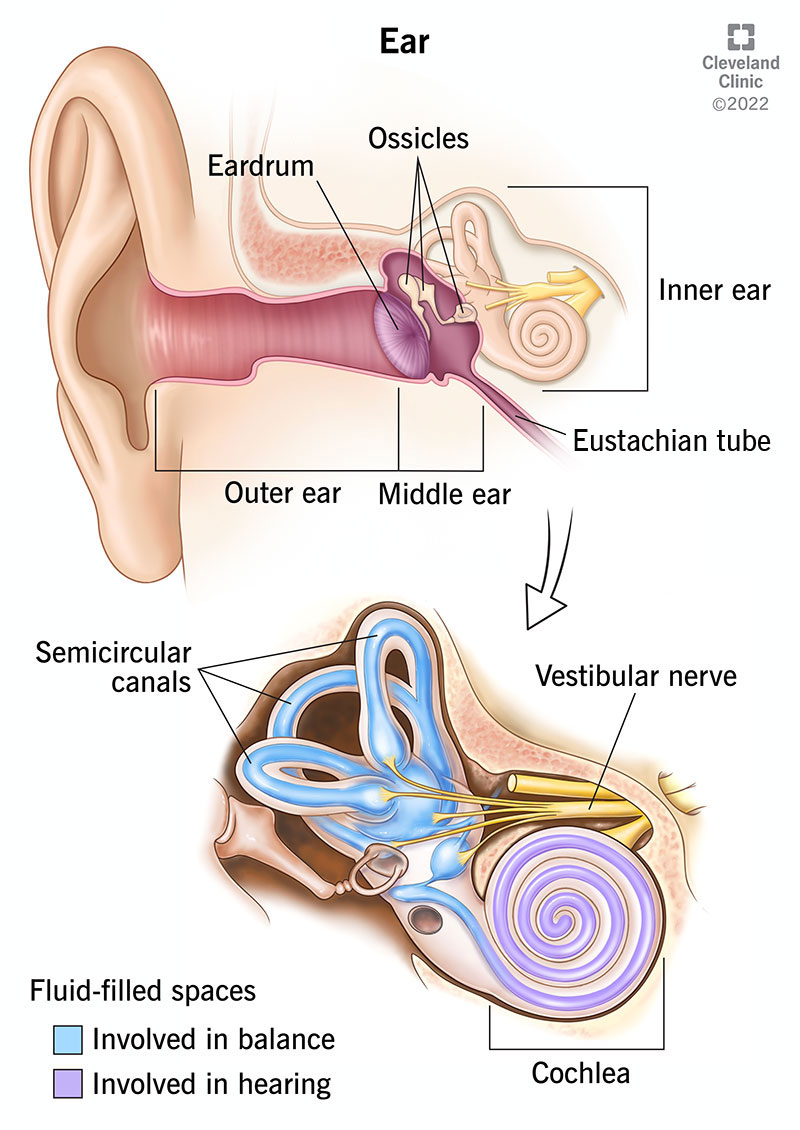
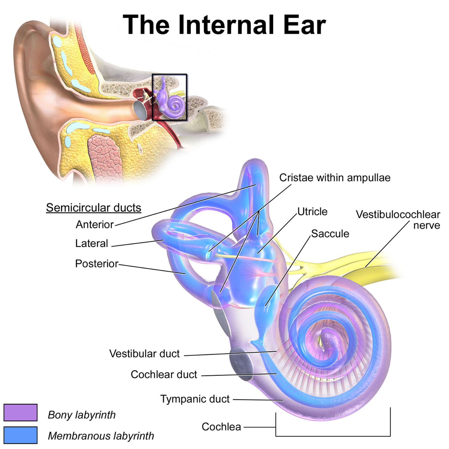
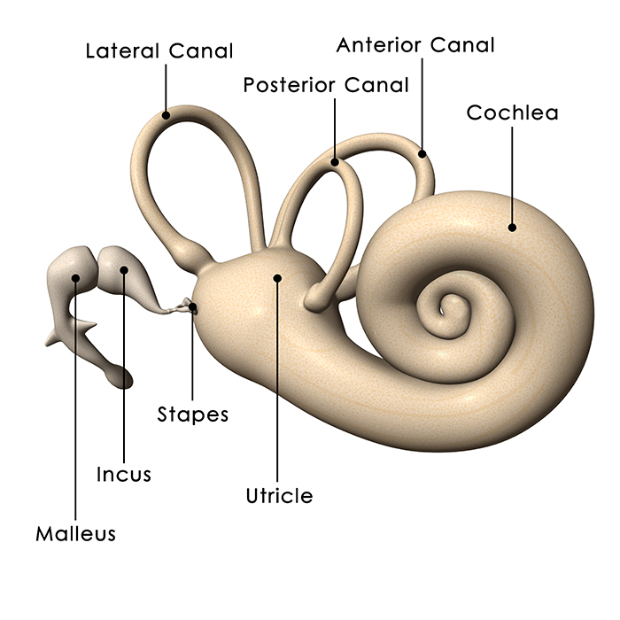
Anatomy What are the parts of the inner ear? Your inner ear has three main parts: your cochlea, semi-circular canals (labyrinth) and your vestibule. Your cochlea supports your hearing and your vestibule and semi-circular canals support your balance. What is the cochlea?
Ear Anatomy Causes of Hearing Loss Hearing Aids Audiology
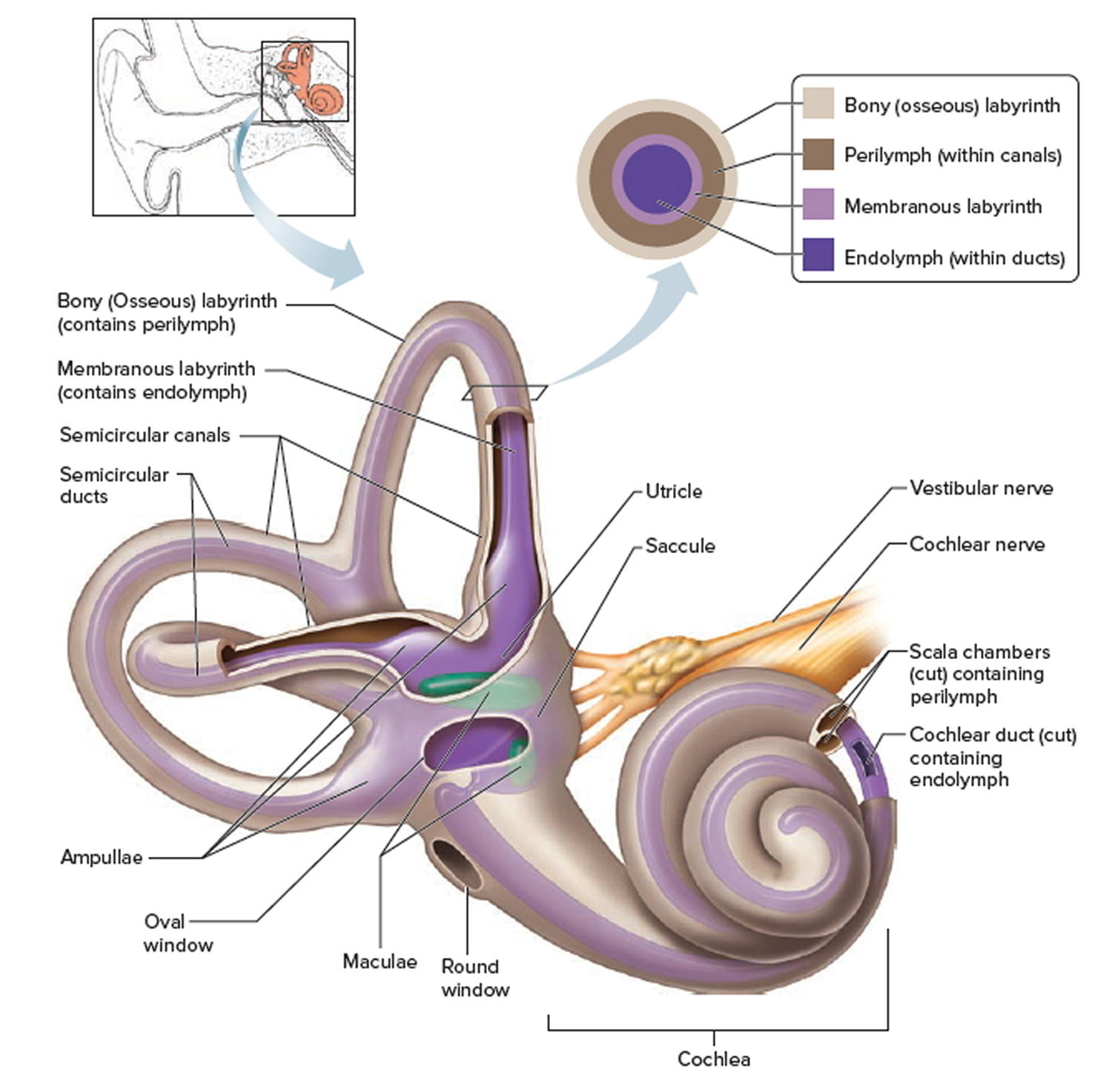
Figure 31.4. Human inner ear anatomy. The inner ear has two structures: the semicircular canals and the spiral-shaped cochlea. The structures of the inner ear are filled with fluid. They are separated from the air-filled middle ear by two membranes called the oval window and the round window. Cross Section of the Cochlea

EarQ Anatomy of the Ear Chart Human ear, Inner ear diagram, Ear anatomy
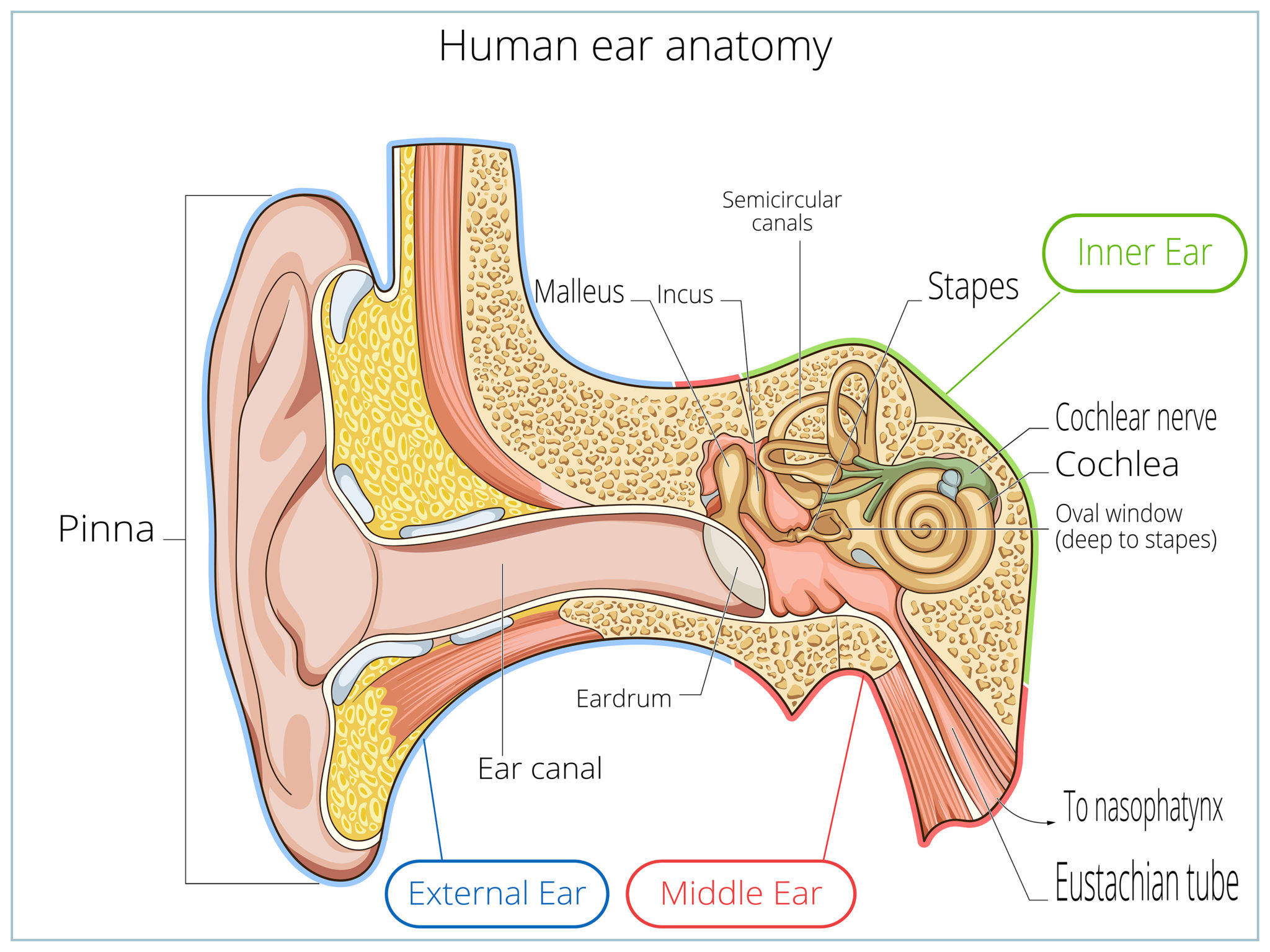
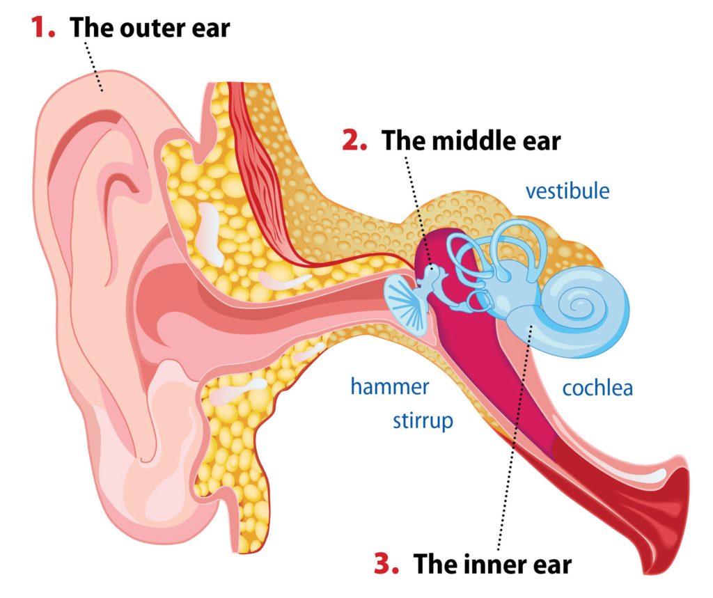
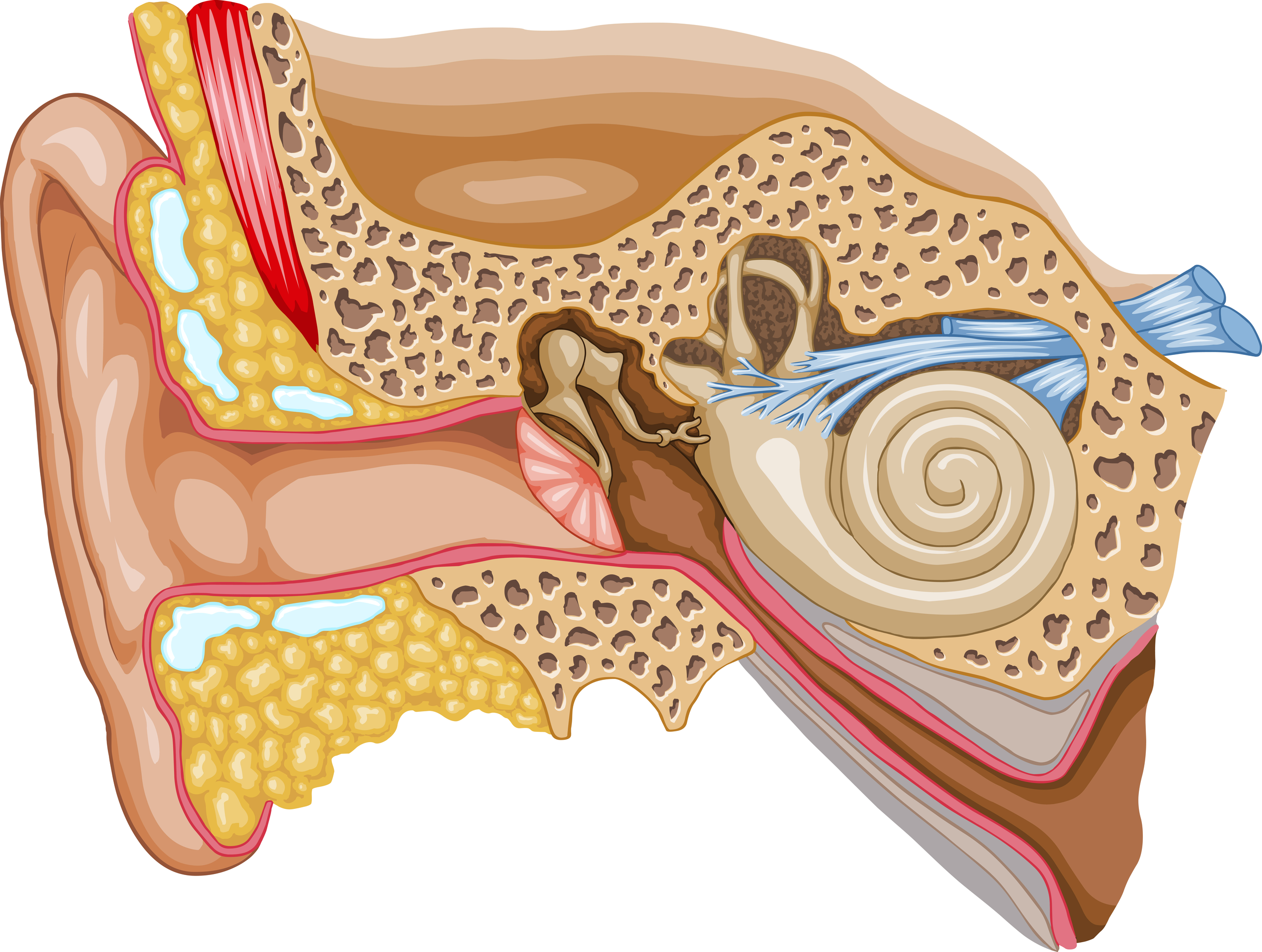
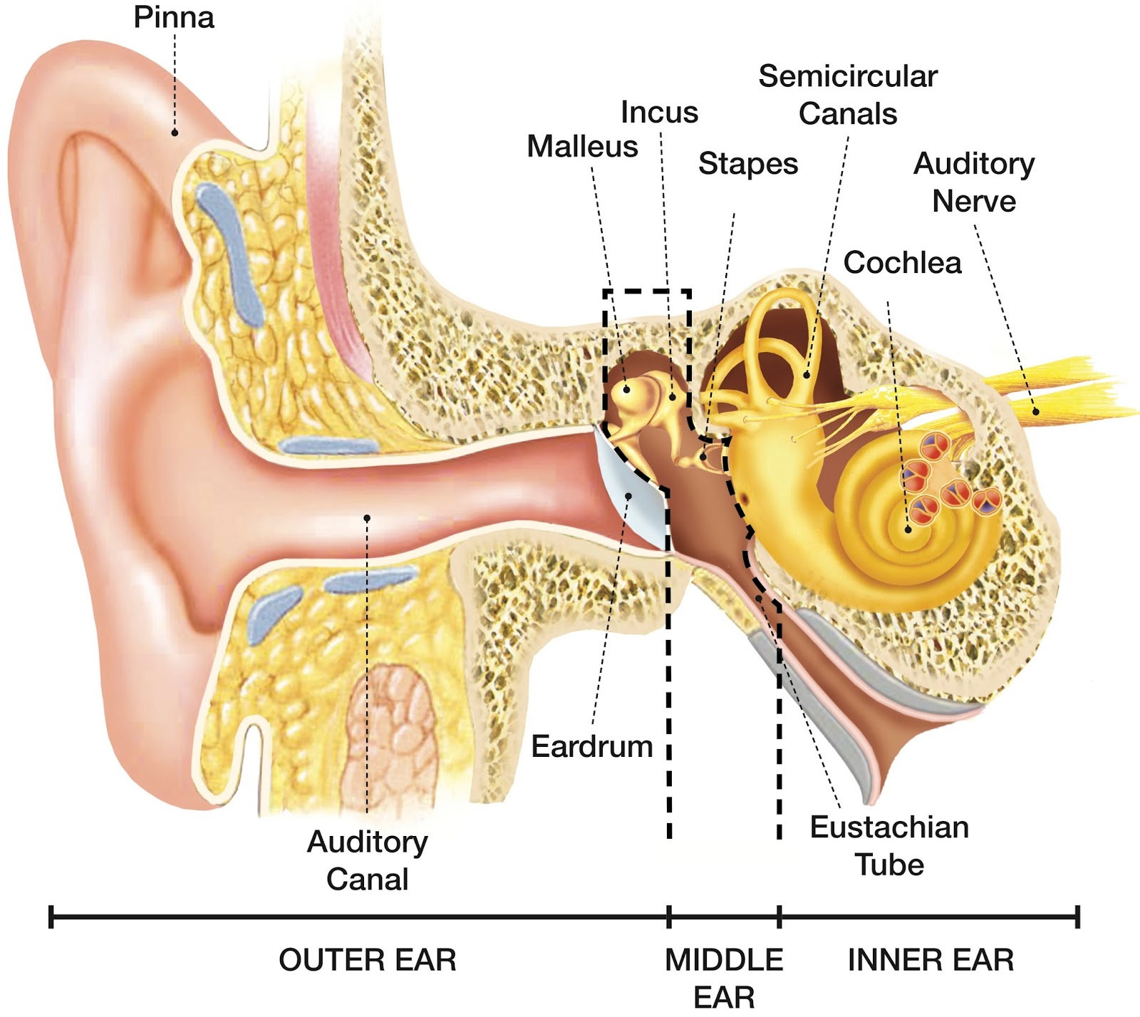
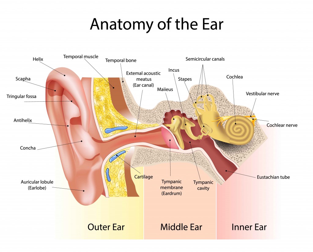
Anatomy Where are my ears located? Your ears are on either side of your head, directly over your temporal lobe. This part of your brain is responsible for hearing, speech, memory and some emotion. Advertisement What are the parts of the ear? The three main parts of your ear include the outer ear, middle ear and inner ear.
How We Perceive Sound Davidson Hearing Aid Centres
human ear, organ of hearing and equilibrium that detects and analyzes sound by transduction (or the conversion of sound waves into electrochemical impulses) and maintains the sense of balance (equilibrium). Understand the science of hearing and how humans and other mammals perceive sound How humans and other mammals perceive sound.
What is conductive hearing loss? Blog of Kiversal
The human ear is the organ of hearing and equilibrium. It detects and analyzes sound by the mechanism of transduction, which is the process of converting sound waves into electrochemical impulses. Audition cannot take place adequately if the anatomy is abnormal. This article will discuss the mechanisms implied in the conduction of sound waves into the ear, and its integration and transmission.
Outer Ear Anatomy Outer Ear Infection & Pain Causes & Treatment
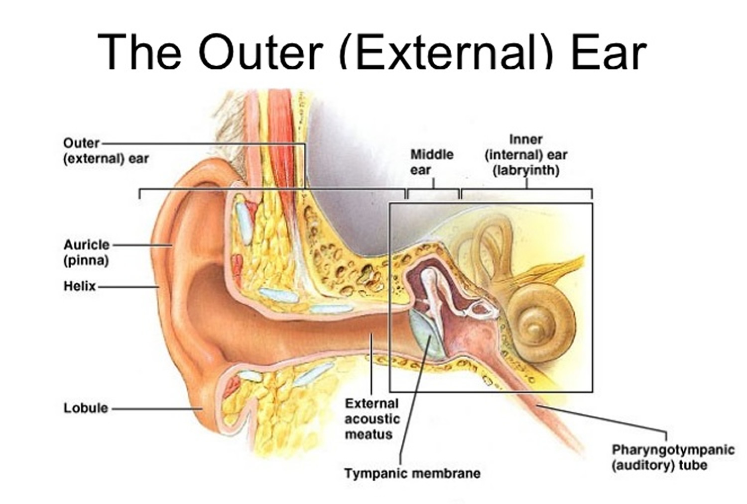
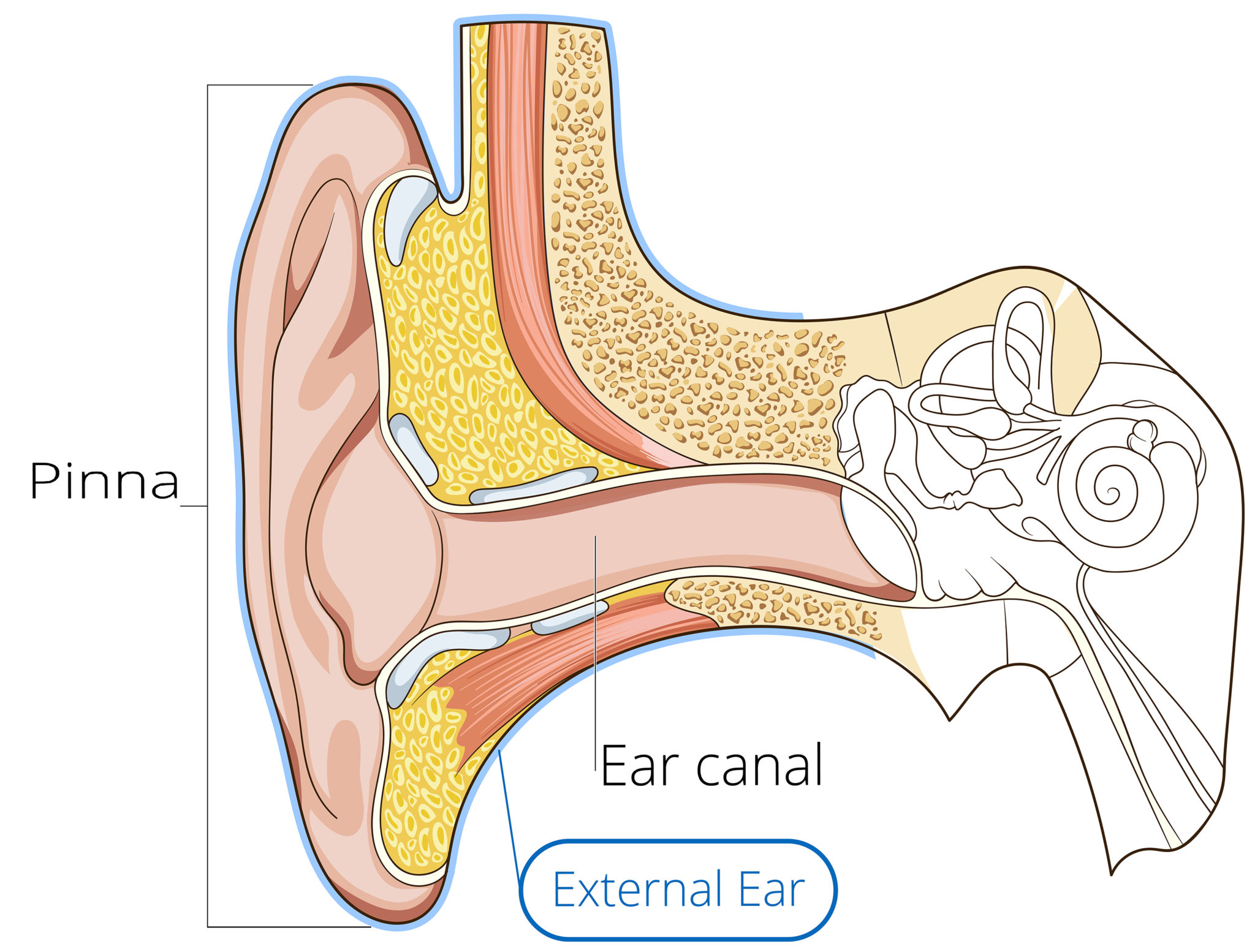
Anatomy and function of the inner ear. The generalized ear of mammals is partitioned into the outer, middle, and inner ears. The outer ear includes the pinna, which funnels sound from the environment into the ear region of the head, and extends from the external surface of the head to the tympanic membrane, or eardrum, via the external acoustic meatus (Fig. 1).

Afbeeldingsresultaat voor middle ear anatomy Ear anatomy, Middle ear
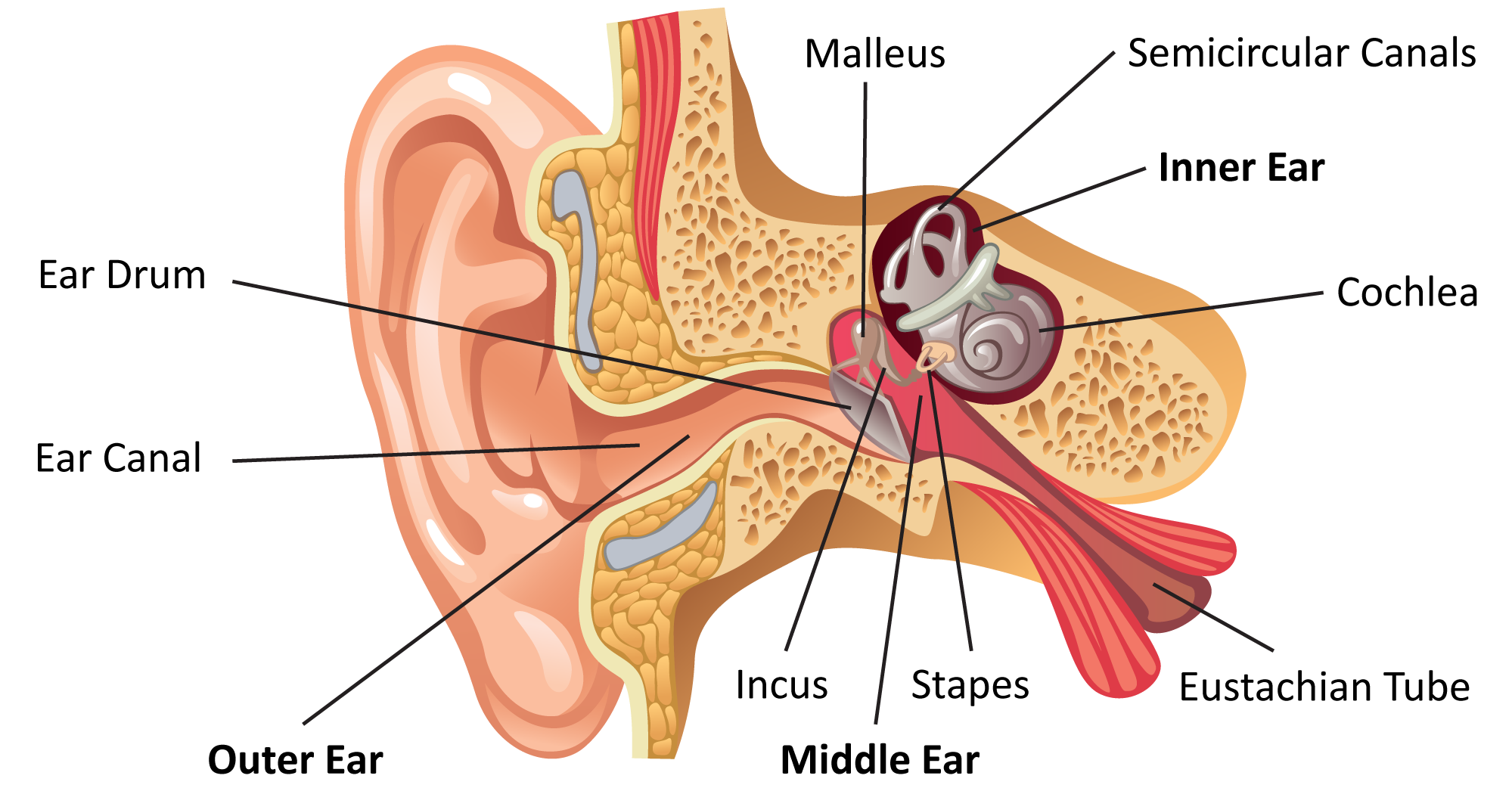
What Is the Anatomy of an Ear? The ear is an unusually complex organ in human anatomy. Don't worry, though—each part has a purpose that is easy to understand. In this section, we describe the anatomy of the ear in simple terms. External Ear Anatomy (Auricle or Pinna)

15.3 Hearing Anatomy & Physiology
The inner ear is located within the bony labyrinth of the temporal bone and contains the cochlea, semicircular canals, utricle, and saccule. These organs make up the membranous labyrinth that is within the bony labyrinth, separated only by perilymph.
Medical problems of the eyes, ears, nose, and throat symptoms
Next to the middle ear in the bone of the skull is a small compartment which contains the hearing and balance apparatus known as the inner ear. The inner ear has two main parts. The cochlea , which is the hearing portion, and the semicircular canals is the balance portion.
Inner Ear Problems Causes & Treatment of inner ear Dizziness & Vertigo
The inner ear is found in the petrous part of the temporal bone between the middle ear laterally, and the internal acoustic meatus medially. It is a small and important area which houses the irregularly shaped vestibulocochlear organ, which kind of looks like a snail shell attached to a few bony rings. Now, the inner ear contains the bony.
SPEECH LANGUAGE PATHOLOGY & AUDIOLOGY HEARING DISORDERS OF THE OUTER EAR
Fig 1 - Overview of the ear Anatomical Position and Structure The inner ear is located within the petrous part of the temporal bone. It lies between the middle ear and the internal acoustic meatus, which lie laterally and medially respectively. The inner ear has two main components - the bony labyrinth and membranous labyrinth.
Inner Ear Discovery Helps Explain How Sound Waves Brain Signals
The inner ear is embedded within the petrous part of the temporal bone, anterolateral to the posterior cranial fossa, with the medial wall of the middle ear, the promontory, serving as its lateral wall.

Ear Anatomy Vestibular Disorders Association
Inner and Middle Ear. The cochlea is the most critical component of the inner ear. It is divided into three fluid-filled chambers, called scalae, that spiral around a bony core. The scala media.
Ear Anatomy Causes of Hearing Loss Hearing Aids Audiology
The inner ear, or labyrinth, is the deepest part of the ear. It is located at the end of the ear canals, resting in a cavity in the temporal bone. The inner ear consists of three parts:.
Human Ear Anatomy Parts of Ear Structure, Diagram and Ear Problems
Internal ear This mixture of bones, nerves, vessels, membranes, and muscles that make up the ear will be described in this article. Contents External ear Auricle External acoustic meatus Tympanic membrane Muscles of the external ear Vasculature of the external ear Innervation of the external ear Middle ear Tympanic cavity Auditory ossicles
We Finally Know Why There's a Bizarre Structure in Our Inner Ears
inner ear, part of the ear that contains organs of the senses of hearing and equilibrium. The bony labyrinth, a cavity in the temporal bone, is divided into three sections: the vestibule, the semicircular canals, and the cochlea.